Revolutionary Technique Makes Mouse Skin See-Through for Brain Imaging
A groundbreaking, reversible method to make mouse skin transparent is transforming brain imaging, offering a new window into neurodevelopment.

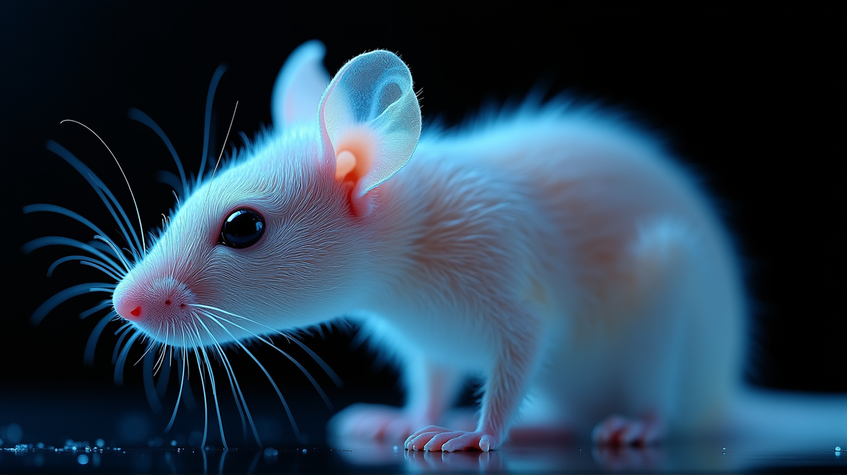
In a stunning breakthrough in the field of neurodevelopmental research, scientists have developed a reversible technique enabling the skin of juvenile mice to become transparent to visible light. This innovation, thoroughly chronicled in the Proceedings of the National Academy of Sciences (PNAS), spreads new light on the already intricate endeavor of brain imaging, revealing neural development over time without invasive procedures or permanent tissue alterations.
Matching Optical Properties
The key challenge in imaging through skin lies in light scattering, a phenomenon that occurs when light traverses materials with differing optical properties, such as water, lipids, and proteins. This mismatch has traditionally disrupted light passage, making it near impossible to observe intricate brain structures beneath the skin. “From a physics perspective, we’re basically a bag of water with biomaterials,” Dr. Mark Brongersma stated, encapsulating the fundamental vision behind this study.
The ingenious approach deployed by Stanford University researchers centers on addressing this discrepancy by augmenting the refractive index of water in the skin tissue. Using a compound called ampyrone, they aligned the refractive index of the treated skin closer to that of surrounding biomaterials, thereby allowing light to pass through with minimal deformation.
The Science Behind Transparency
Ampyrone, renowned for its ability to absorb ultraviolet light while permitting visible light transmission, plays a crucial role in this transformation. The technique adopts the refractive index as a fundamental parameter to control light bending through different materials, thereby simplifying the complexity of light scattering through biological tissues.
This landmark method not only achieves transparency without damaging the skin or skull, but its effects wane over time, implying the prospect for repeated applications—a significant leap from conventional imaging strategies limited by wavelength specificity and invasiveness.
Illuminating Brain Activity
Unlike preceding techniques necessitating invasive procedures or closely tied to singular wavelengths, this novel method embraces the entire visible spectrum. It enables scientists to capitalize on fluorescent protein markers such as green and yellow fluorescence, which are pivotal in depicting neural activity. “This opens a literal window to peek into the brain’s development,” Dr. Guosong Hong remarked, emphasizing the potential of this advancement.
The natural translucence of juvenile mouse skulls supports sending and receiving signals from neurons visible through the skull up to four weeks of age. Utilizing this method, researchers surveyed neuronal structures and activity patterns in both tranquilized and conscious mice, unveiling how brain activity corresponds to specific sensory stimuli like directional air puffs at whiskers.
Implications for Future Research
This transparent skin methodology promises to bolster the scope and depth of neurodevelopmental research, offering an unprecedented opportunity to chronicle how neuronal circuits shape and transform throughout early life. The innovative approach facilitates repeated imaging over extended periods, rendering researchers capable of detecting subtle transformations in brain structure and function.
Building upon prior efforts, which leveraged different compounds for imaging internal organs using red light, this ampyrone-infused protocol pushes the boundaries of observable biological markers to embrace the full spectrum of visible light. According to Technology Networks, it’s an evolving scene that undoubtedly excites and inspires future endeavors in neurological disorder studies, paving smoother paths toward understanding our most complex organ—the brain.